Topographic idiosyncrasies
Different brains encode the same information in distinct ways Haxby et al., 2020. Across individuals, the same brain function may be implemented in different anatomical locations, while the same anatomical region may support different functions. These idiosyncrasies in the correspondence between function and anatomy---often referred to as topographic idiosyncrasies---pose a major challenge for studying brain function across individuals.
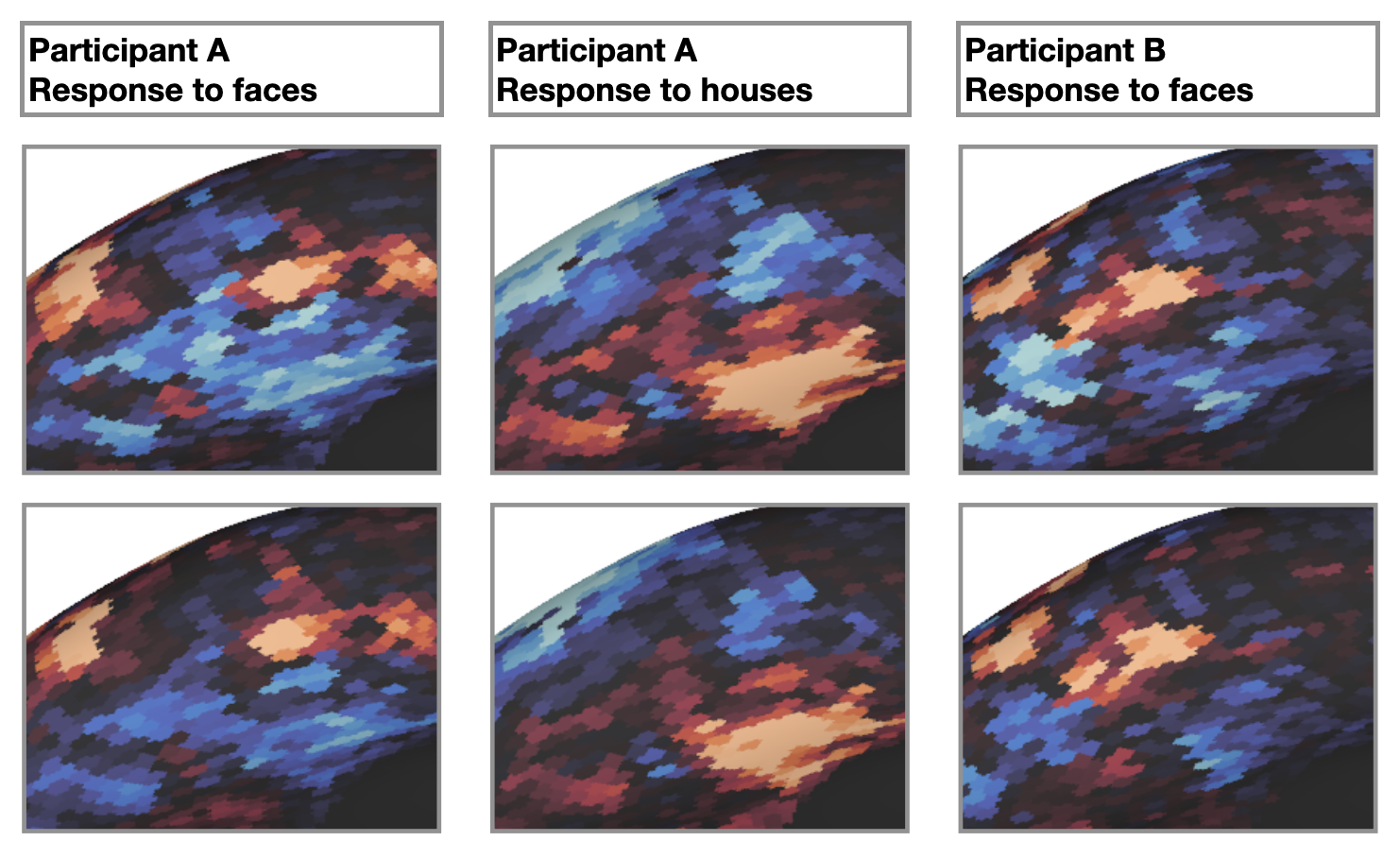
Fig 1. Topographic idiosyncrasies. Within each brain, response patterns to a given stimulus are reliable across independent measurements (top and bottom rows). The brain shows distinct response patterns to different stimuli (left and middle columns). However, across brains, response patterns to the same stimulus differ (left and right columns). See Feilong & Zhang (2025) for a discussion of how these idiosyncrasies can be resolved
Predicting individualized brain responses
To study these functional regions, rather than anatomical locations, researchers often use functional localizers to identify these regions in each individual brain. However, acquiring such data can be challenging in many studies. Hyperalignment Haxby et al., 2020, a functional alignment method, provides a powerful solution to this problem. Hyperalignment establishes functional correspondence across individuals, allowing for projecting fMRI data from one brain to another. The projected data, particularly when aggregated across multiple source individuals, can serve as an accurate prediction of the target individual’s brain responses to the same stimuli Jiahui et al., 2020Jiahui et al., 2023Feilong et al., 2023Guntupalli et al., 2016Haxby et al., 2011.
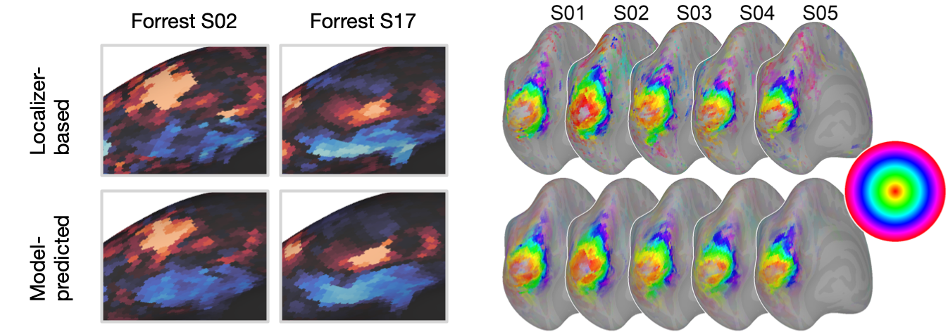
Fig 2. Predicting individualized brain responses using the INT model. The top row shows the face-selectivity map and eccentricity map based on the localizer scans. The bottom row shows the predicted maps using the Individualized Neural Tuning (INT) model Feilong et al., 2023. Note that the localizer-based maps are not noise-free, and the differences between the predicted and localizer-based maps partly reflect noise in the localizer data.
References
- Haxby, J. V., Guntupalli, J. S., Nastase, S. A., & Feilong, M. (2020). Hyperalignment: Modeling shared information encoded in idiosyncratic cortical topographies. Elife, 9, e56601. https://doi.org/10.7554/eLife.56601
- Feilong, M., & Zhang, Y. (2025). Advancing neural decoding with deep learning: Computational neuroscience. Nature Computational Science, 1–2. https://doi.org/10.1038/s43588-025-00837-2
- Jiahui, G., Feilong, M., di Oleggio Castello, M. V., Guntupalli, J. S., Chauhan, V., Haxby, J. V., & Gobbini, M. I. (2020). Predicting individual face-selective topography using naturalistic stimuli. NeuroImage, 216, 116458. https://doi.org/10.1016/j.neuroimage.2019.116458
- Jiahui, G., Feilong, M., Nastase, S. A., Haxby, J. V., & Gobbini, M. I. (2023). Cross-movie prediction of individualized functional topography. Elife, 12, e86037. https://doi.org/10.7554/eLife.86037
- Feilong, M., Nastase, S. A., Jiahui, G., Halchenko, Y. O., Gobbini, M. I., & Haxby, J. V. (2023). The individualized neural tuning model: Precise and generalizable cartography of functional architecture in individual brains. Imaging Neuroscience, 1, 1–34. https://doi.org/10.1162/imag_a_00032
- Guntupalli, J. S., Hanke, M., Halchenko, Y. O., Connolly, A. C., Ramadge, P. J., & Haxby, J. V. (2016). A model of representational spaces in human cortex. Cerebral Cortex, 26(6), 2919–2934. https://doi.org/10.1093/cercor/bhw068
- Haxby, J. V., Guntupalli, J. S., Connolly, A. C., Halchenko, Y. O., Conroy, B. R., Gobbini, M. I., Hanke, M., & Ramadge, P. J. (2011). A common, high-dimensional model of the representational space in human ventral temporal cortex. Neuron, 72(2), 404–416. https://doi.org/10.1016/j.neuron.2011.08.026